New Product Launch | T-Cell Expansion Medium (Serum-Free)
Publication Date:2024-06-05
Foreword
T cells are a type of white blood cell in the human immune system primarily responsible for cellular immunity within the body. They play a crucial role in combating viral infections, tumors, and other pathogens. Traditional T-cell culture media typically contain fetal bovine serum or other serum components derived from animal sources. However, these serums may present issues such as batch-to-batch variability, unknown factors, and potential infection risks. Developing serum-free media reduces variables in the culture process, enhances reproducibility and standardization, and avoids ethical and regulatory concerns associated with animal serum use. This aligns with the sustainable and ethical trends in modern biotechnology.
Product Introduction
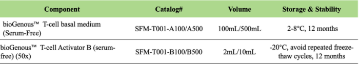
bioGenous™ T-Cell Expansion Medium (Serum-Free) is a specially formulated cell culture medium designed for the in vitro culture and efficient expansion of human T lymphocytes without the need for added serum. This serum-free T-cell expansion medium contains complete nutrients essential for T-cell growth, supporting T-cell expansion from peripheral blood mononuclear cells (PBMCs) and is suitable for reactivated T-cell expansion cultures. Expanded T-cells are applicable for various in vitro studies, including T-cell assays for drug activity detection and screening, as well as T-cell co-culture experiments with tumor organoids.
Product Features
Serum-free, xeno-free, and exogenous growth factor-free
Supports in vitro activation, expansion, and high-density culture of T cells
Exhibits similar phenotype and function to T cells cultured in serum-containing media
Maintains a CD4:CD8 ratio comparable to baseline after expansion
Requires no magnetic bead activation for T cells
Product Data Presentation
PBMC In Vitro Proliferation:
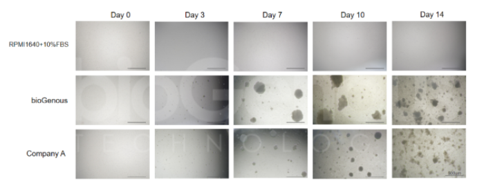
Figure 1 Comparison of in vitro growth states of PBMCs isolated from patients
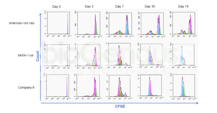
Figure 2CFSE staining analysis comparing the in vitro proliferation of PBMCs isolated from patients.
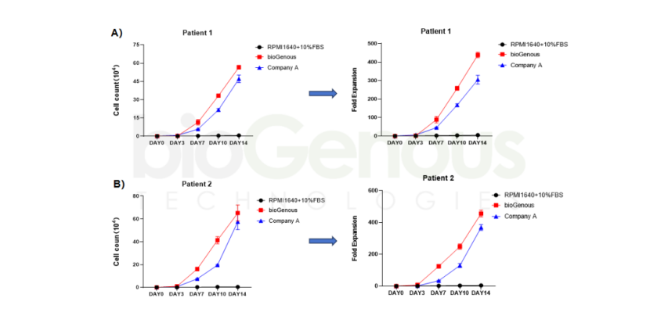
Figure 3 Comparison of in vitro expansion of PBMCs isolated from different patients: A) Trends in cell number and amplification multiples of PBMCs isolated from the first patient during in vitro expansion; B) Trends in the number and amplification multiples of in vitro expanded PBMCs isolated from the second patient.
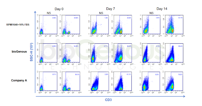
In vitro proliferation of T cells in PBMCs:
Figure 4In vitro expansion of T cells from PBMCs isolated from patients
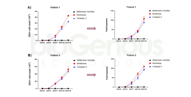
Figure 5 Comparison of T-cell in vitro expansion from PBMCs isolated from different patients: A) Trends in T-cell in vitro expansion numbers and amplification multiples from PBMCs isolated from the first patient; B) Trends in T-cell in vitro expansion numbers and amplification multiples from PBMCs isolated from the second patient.
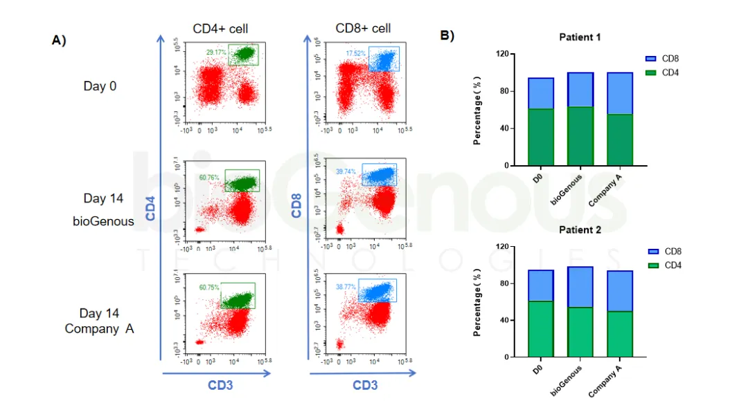
Marker Expression in T Cells Following In Vitro Proliferation in PBMCs:
Figure 6 Comparison of CD4 and CD8 Expression in PBMCs Before and After In Vitro Expansion from Different Patient Sources: A) Proportion of CD4+ and CD8+ Cells in PBMCs; B) Proportion of CD4+ and CD8+ Cells in CD3+ Cells